Figure 3.

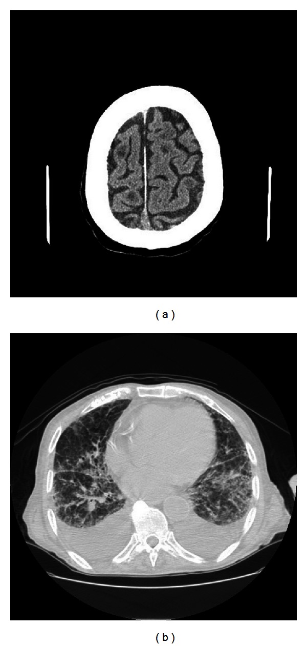
(a) Computerized tomography (CT) scan of the patient's brain showing multiple enhancing focal lesions. (b) Chest CT scan showing multiple nodular lesions and bilateral pleural effusion.

(a) Computerized tomography (CT) scan of the patient's brain showing multiple enhancing focal lesions. (b) Chest CT scan showing multiple nodular lesions and bilateral pleural effusion.