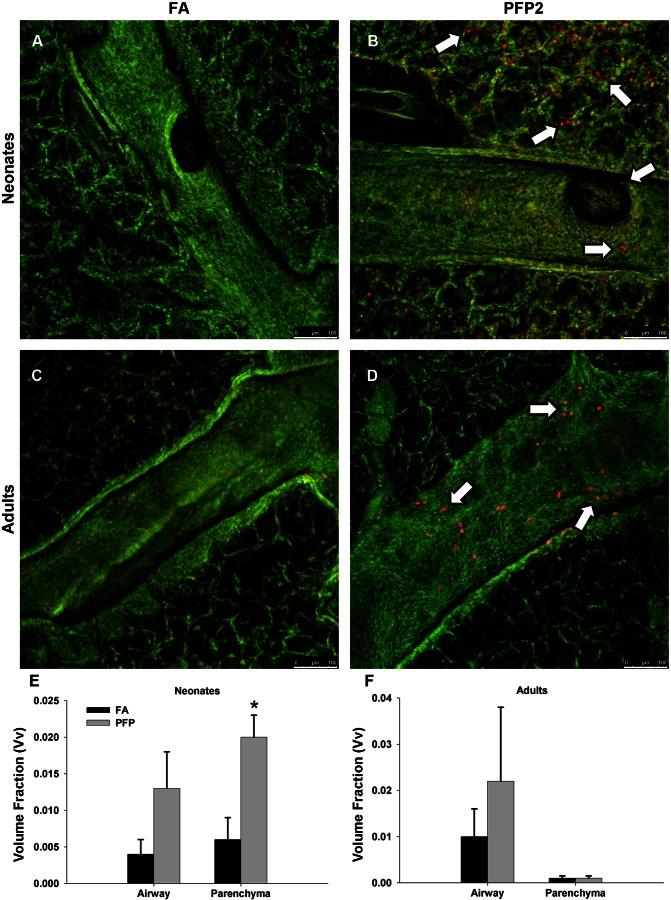
Fig. 2.
Airway cellular toxicity following PFP exposure. In situ ethidium homodimer-1 staining was analyzed using confocal microscopy in 7-day postnatal (A and B) and adult (C and D) rats exposed to either filtered air (FA, A and C) or PFP2 (B and D). Overall, ethidium staining was scarce in either 7-day-old neonates (A) or adults (C) reared in FA. Ethidium-positive cells (white arrows) were observed distributed in both bronchiolar epithelium and parenchyma in neonatal animals (B). Comparatively, the majority of cytotoxic cells was present in bronchiolar airways and was virtually absent in the parenchyma (D). Scale bars are 100 μm. Stereological quantification revealed an increased volume fraction of ethidium homodimer-1-positive cells after exposure. The volume fraction of ethidium-positive cells of neonates increased in both airway and parenchyma after exposure, but this increase was significant only in the parenchyma (E). Adult volume fractions also trended higher in the airways but were unaffected by PFPs in the parenchyma (F). Data are plotted as means ± SE (n = 4–5 rats/group, per compartment). *P < 0.05, significantly different from FA in the same compartment.

