Fig. 3.
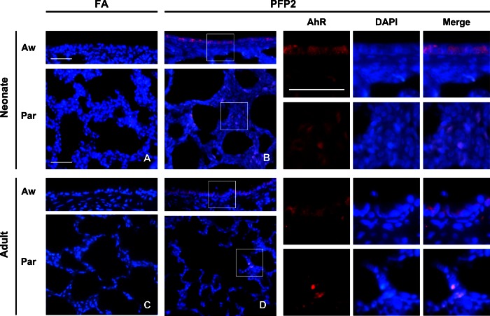
AhR immunofluorescence. AhR immunofluorescence (red) was overlaid upon DAPI nuclear (blue) staining in neonate (A and B) and adult (C and D) lungs exposed to either FA (A and C) or PFP2 (B and D). AhR staining was virtually absent in FA-treated animals of either age. However, robust AhR staining was observed in both airway and parenchyma compartments in both ages. High-magnification insets are denoted by white squares and separate images showing AhR and DAPI colocalization, presented adjacent to B and D, respectively. Aw, airway; Par, parenchyma.

