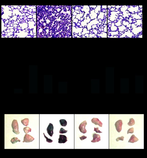
Fig. 8.
Histological assessment of the effect of gVPLA2 inhibition on ventilator-induced lung injury and analysis of lung vascular leak. Wild-type littermate control (pla2g5+/+) with or without injection with gVPLA2 blocking antibody or gVPLA2 knockout (pla2g5−/−) were exposed to HTV. Spontaneously breathing animals were used as controls. A: histological analysis of lung tissue (×40 magnification). Whole lungs (4 to 6 animals from each experimental group) were agarose inflated in situ, fixed with 10% formalin, and used for histological evaluation by hematoxylin and eosin staining as described in materials and methods. B: protein concentration in BAL fluid from HTV-exposed wild-type littermate control (pla2g5+/+) mice with or without injection with gVPLA2 blocking antibody or gVPLA2 knockout (pla2g5−/−) mice. C: lung vascular leak was assessed by measurements of Evans blue leakage into lung tissues, as described in materials and methods. Quantitative analysis was performed by spectrophotometric measurement of Evans blue extracted from the lung tissue samples. The data represent the means ± SE of 6 samples; *P < 0.05 vs. HTV alone in wild-type mice. D: images of lung preparations depicting Evans blue accumulation in the lung tissues.

