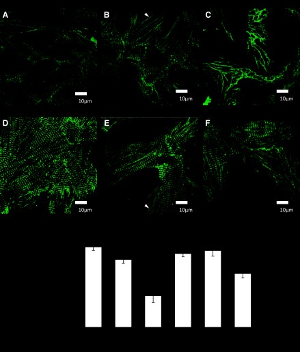
Fig. 1.
Immunostaining of cultured neonatal rat myocytes for α-actinin (green). A: control myocytes. B: myocytes following 10% mechanical stretch for 48 h. C: myocytes following 20% stretch. D: myocytes treated with nitro-l-arginine methyl ester (l-NAME) for 48 h. E: myocytes treated with l-NAME and 10% stretch F: myocytes treated with l-NAME and 20% stretch. White arrows show areas lacking in sarcomeres. G: quantitation of intact sarcomeres in cultured neonatal myocytes (P < 0.05, compared with control; n ≥ 5 cultures). Scale bars = 10 μm.

