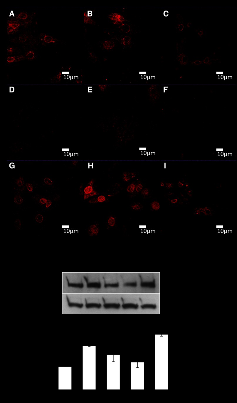
Fig. 6.
Immunostaining of neonatal myocytes to show the subcellular localization of the three nitric oxide synthase isoforms in cardiac myocytes. A, D, and G: myocytes immunostained for endothelial (eNOS), neuronal (nNOS), and inducible (iNOS) forms, respectively, in control cells. B, E, and H: cells after 10% stretch for 48 h at 1 Hz. C, F, and I: images following 20% stretch for 48 h at 1 Hz. Scale bars = 10 μm. J: Western blot of LPP protein following 48-h treatments with different NOS inhibitors: 5 mM l-NAME (a nonspecific NOS inhibitor), 10 μM of the iNOS inhibitor 1400W, and 10 μM of the nNOS inhibitor N-{(4S)-4-amino-5-[(2-aminoethyl)amino]pentyl}-N′-nitroguanidine tris(trifluoroacetate) salt and a combination of iNOS and nNOS inhibitors. K: LPP protein expression in response to l-NAME and to separate inhibition of iNOS or nNOS. *P < 0.05, compared with control; n = 3 cultures.

