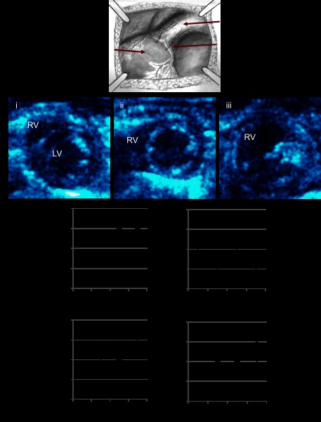
Fig. 1.
A: pulmonary insufficiency was created by entrapping the pulmonary valve leaflets. Echocardiographic functional assessment. Echocardiograms performed during the progression of volume overload demonstrated (B) increasing right ventricular (RV) dilation (i), sham, mild RV dilation at 2 wk (ii), moderate to severe RV dilation at 1, 3, and 6m (iii), increasing heart rate (C), early decrease in right ventricular outflow tract (D; RVOT) shortening fraction followed by normalization, an early reversal in the tricuspid valve early and late diastolic filling velocities (E/A ratio) followed by pseudonormalization (E), and no change in the RV free wall velocity (S' peak; F). PI, pulmonary insufficiency; LV, left ventricular. *P < 0.05 vs. sham.

