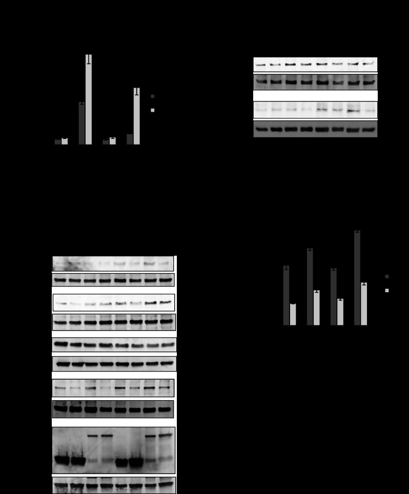
Fig. 2.
PHB-induced ARE activation, NQO-1 and HO-1 protein expression, and decreased intracellular reactive oxygen species (ROS) levels during TNFα treatment are not exclusively mediated via Nrf2. A: relative ARE-driven luciferase activity in Caco-2-BBE cells cotransfected with pEGFPN1-PHB (PHB) or pEGFPN1-vector (V) and Nrf2 small interfering RNA (siNrf2) or negative control small interfering RNA (siRNA, TNFα). Values are means ± SE (n = 6 per treatment). *P < 0.05, **P < 0.01. B: Western blot of NQO-1, HO-1, and endogenous PHB in cells transfected and treated as described in A. Duplicate blots were probed with anti-GFP or anti-Nrf2 to ensure GFP-PHB transfection efficiency and Nrf2 knockdown, respectively. β-Actin was used as a loading control. Blots are representative of 3 independent experiments. C: Western blot of Nrf2 protein expression in nuclear and cytosolic extracts. Histone H3 and β-actin were used as loading controls for nuclear or cytosolic extracts, respectively. Blots are representative of 3 independent experiments. D: 2′,7′-dichlorofluorescein (DCF) fluorescence in Caco-2-BBE cells. RFU, relative fluorescence units. Values are means ± SE (n = 8 per treatment). *P < 0.05, **P < 0.01.

