
Fig. 6.
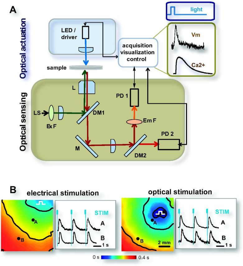
All-optical actuation, sensing, and control of cardiac function. A: system for all-optical contactless cardiac electrophysiology, built around a microscope. Optical actuation is achieved through collimated light-emitting diode (LED)-produced light; optical sensing of voltage (Vm) and calcium (Ca2+) is done by voltage-sensitive dye (di-8-ANEPPS) and calcium-sensitive dye (Rhod-4), respectively; shown are actual records (light pulse was 50 ms, 0.2 mW/mm2). Computer controls the LED driver and the acquisition by the photodetectors (PD1, PD2), thus allowing for a closed-loop feedback control. L, objective lens; LS, light source for imaging; Ex F and Em F, excitation and emission filters, respectively; M, DM1, and DM2, full and dichroic mirrors, respectively. B: all-optical interrogation of cardiac function over time and space by combining high-resolution optical mapping with optogenetic actuation [used with permission from Jia et al. (64)]: waves of excitation in cardiac monolayers, triggered by electrical and optical pacing at 0.5 Hz, and captured by activation maps. Color represents time of activation; isochrones are shown in black at 0.15 s. Calcium transients (Rhod-4) in response to electrical or optical stimulation are shown from two locations, normalized fluorescence. Blue marks indicate time of stimulation (electrical pulses were 10 ms; and optical, 20 ms each). In A and B, cell delivery approach of ChR2 was used.