
Figure 2.
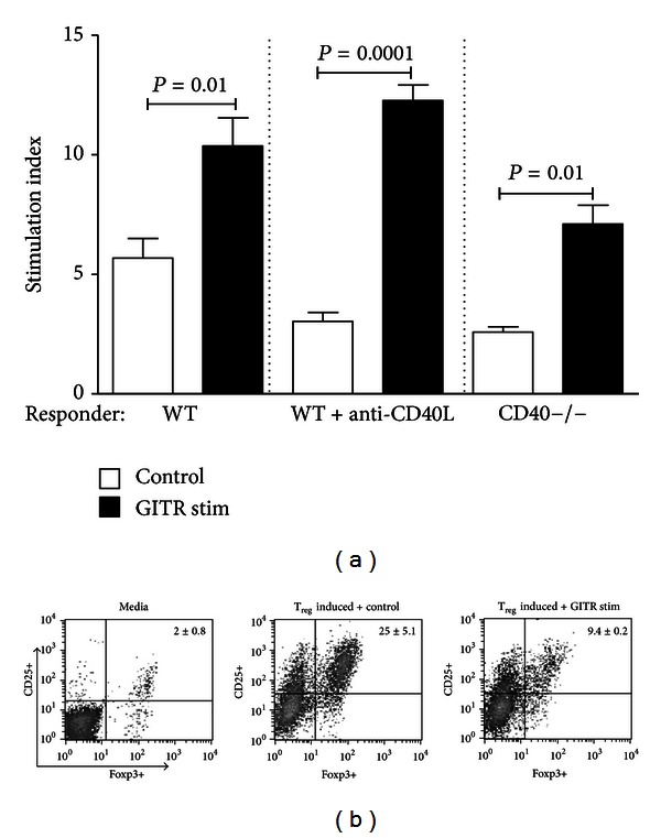
GITR stimulation increases graft-reactive Teff cell proliferation and inhibits Treg development. (a) WT C57BL/6 splenocytes were cultured in the presence of irradiated WT BALB/c splenocytes (WT) or with irradiated WT BALB/c splenocytes + 100 μg/mL anti-CD40L mAb (WT+anti-CD40L); CD40−/− C57BL/6 splenocytes were cultured with irradiated CD40−/− BALB/c splenocytes. Cultures were maintained in the presence of 100 μg/mL of anti-GITR mAb (black bars) or control IgG (open bars) for 5 days and were assayed for proliferation by [3H] thymidine incorporation. Stimulation index was defined as responder + stimulator cpm/responder alone cpm. Average values generated from 3 independent experiments are depicted, and significance was determined via paired Student's t-test. (b) Naïve splenocytes were isolated from Foxp3-GFP knock-in mice and were cultured for 72 hours in media (left panel), in media containing 10 U/mL recombinant IL-2, 10 ng/mL recombinant TGF-β, and 2% anti-CD3 mAb (Treg inducing media) + 100 μg/mL IgG isotype control (middle panel), or in Treg inducing media + 100 μg/mL anti-GITR mAb (right panel). Examples of CD25+ versus Foxp3 GFP+ cell staining from 3 independent experiments are pictured along with the average % staining ± SEM.