
Figure 1.
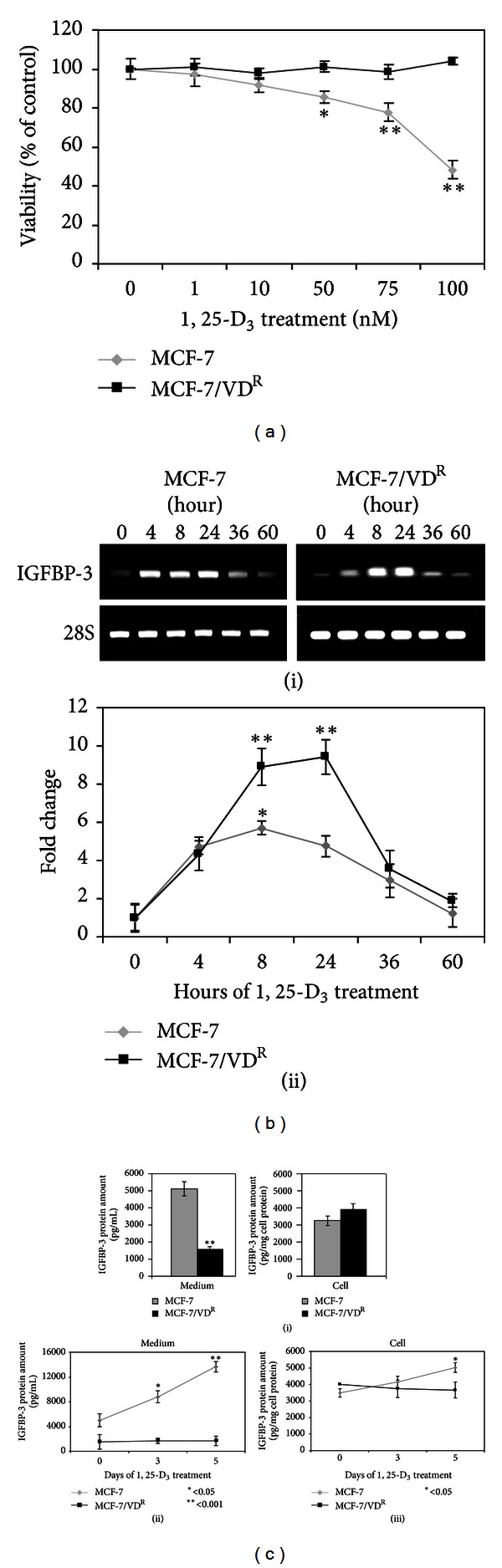
Effect of 1, 25-D3 on MCF-7 and MCF-7/VDR cell viability and IGFBP-3 expression. (a) MCF-7 and MCF-7/VDR cells were treated with increasing concentrations of 1, 25-D3 (up to 100 nM) or 0.1% ethanol vehicle as a control for 6 days. Cell viability was determined by neutral red assay. Means of 3 separate experiments are shown. *P < 0.05 and **P < 0.001 are statistically significant compared to the control. (b) (i) MCF-7 and MCF-7/VDR cells were treated with 100 nM 1, 25-D3 for up to 60 hours. IGFBP-3 mRNA expression was measured by RT-PCR. 28S mRNA expression was used as house-keeping gene. Nontreated cells were used as controls. (ii) Densitometric analysis of of IGFBP-3 mRNA expression. Data shown means of three separate experiments. (c) Intracellular IGFBP-3 levels and secretion into medium was determined by ELISA in MCF-7 and MCF-7/VDR cells. (i) IGFBP-3 expression and secretion into the medium in untreated MCF-7 and MCF-7/VDR cells. (ii) IGFBP-3 secretion into the medium in MCF-7 and MCF-7/VDR cells treated with 100 nM of 1, 25-D3 for up to 5 days quantitated by ELISA. (iii) The amount of intracellular IGFBP-3 production by MCF-7 and MCF-7/VDR cells treated with 100 nM of 1, 25-D3 was measured at day 0, 3, and 5 by ELISA. Means of 3 separate experiments are shown. *P < 0.05 and **P < 0.001 are statistically significant compared to the control.