
Fig. 3.
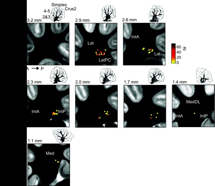
Location of WB and TB. Location are of cells recorded superimposed on photographs of 300-μm sagittal sections of living DCN slices arranged from lateral to medial. RI is indicated by color scale at top right. Drawings at top right of each image show the entire cerebellum at each plane of section. The dorsal-ventral and anterior-posterior axes are indicated. Numbers indicate approximate mediolateral distance of each section from the midline according to Paxinos and Watson (1998). Abbreviations: Lat, lateral nucleus; Lat-PC, lateral nucleus pars compacta; IntA, interpositus anterior; Med, medial nucleus; MedDL, medial nucleus, dorsolateral protuberance.