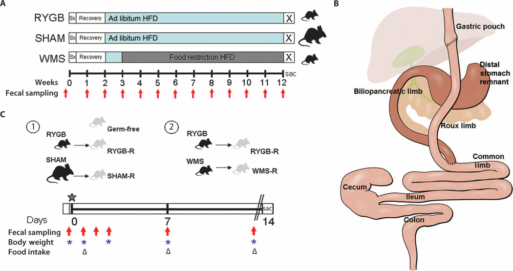
Fig. 1.
Schematic of experimental design. (A) DIO C57BL/6J mice fed a 60% HFD underwent either RYGB or sham operations and 2-week recovery on liquid diet before returning to HFD (blue bars). Sham animals that had successfully regained body weight within 3 weeks after surgery were divided into an ad libitum–fed SHAM group or food-restricted to match the weight of the RYGB animals (WMS). Fresh fecal samples were collected preoperatively and weekly for 12 weeks after surgery for microbiota analysis (red arrows). (B) Graphic of the RYGB anatomy and segments collected for luminal content and mucosal scrapings along the length of the gastrointestinal tract. Segments representative of the RYGB anatomy were also collected in SHAM and WMS animals. (C) Design of microbiota transfer experiments of the cecal contents from a representative donor animal from each group, depicted in (A), into germ-free mice (star), indicating timing of collection of fecal samples for microbiota analysis (red arrows), body weights (blue asterisks), and food intake (triangles). At the end of the colonization period, animals were fasted overnight (double lines), and final body weights, serum metabolic parameters, and adiposity scores were obtained.

