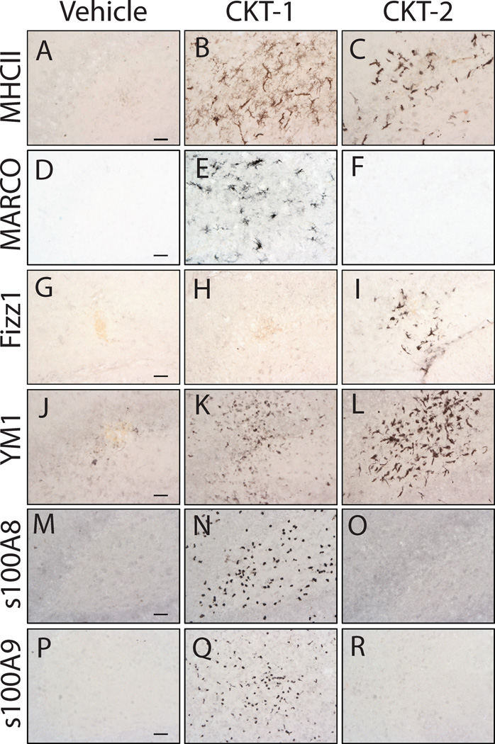
Figure 3.
Photomicrographs of immunohistochemical stains for microglia/ macrophage markers in 6-month-old mice that received cytokine cocktails. Mice were treated intracranially with vehicle (PBS) (Panels A, D, G, J, M, P), CKT-1 (IL-1β, IL-12, TNF-α) (Panels B, E, H, K, N, Q), and CKT-2 (IL-4, IL-13) (Panels C, F, I, L, O, R). Images were centered on the injection sites and stained for MHCII (A–C), MARCO (D–F), FIZZ-1 (G–I), YM1 (J–L), s100A8/ calgranulin A (M–O), and s100A9/ calgranulin B (P–R). MHCII and YM1 were both increased by CKT-1 and CKT-2. Selective induction of MARCO, s100A8 and s100A9 was observed by CKT-1, whereas CKT-2 selectively induced FIZZ1. Scale bar= 25µm.

