Fig. 3.
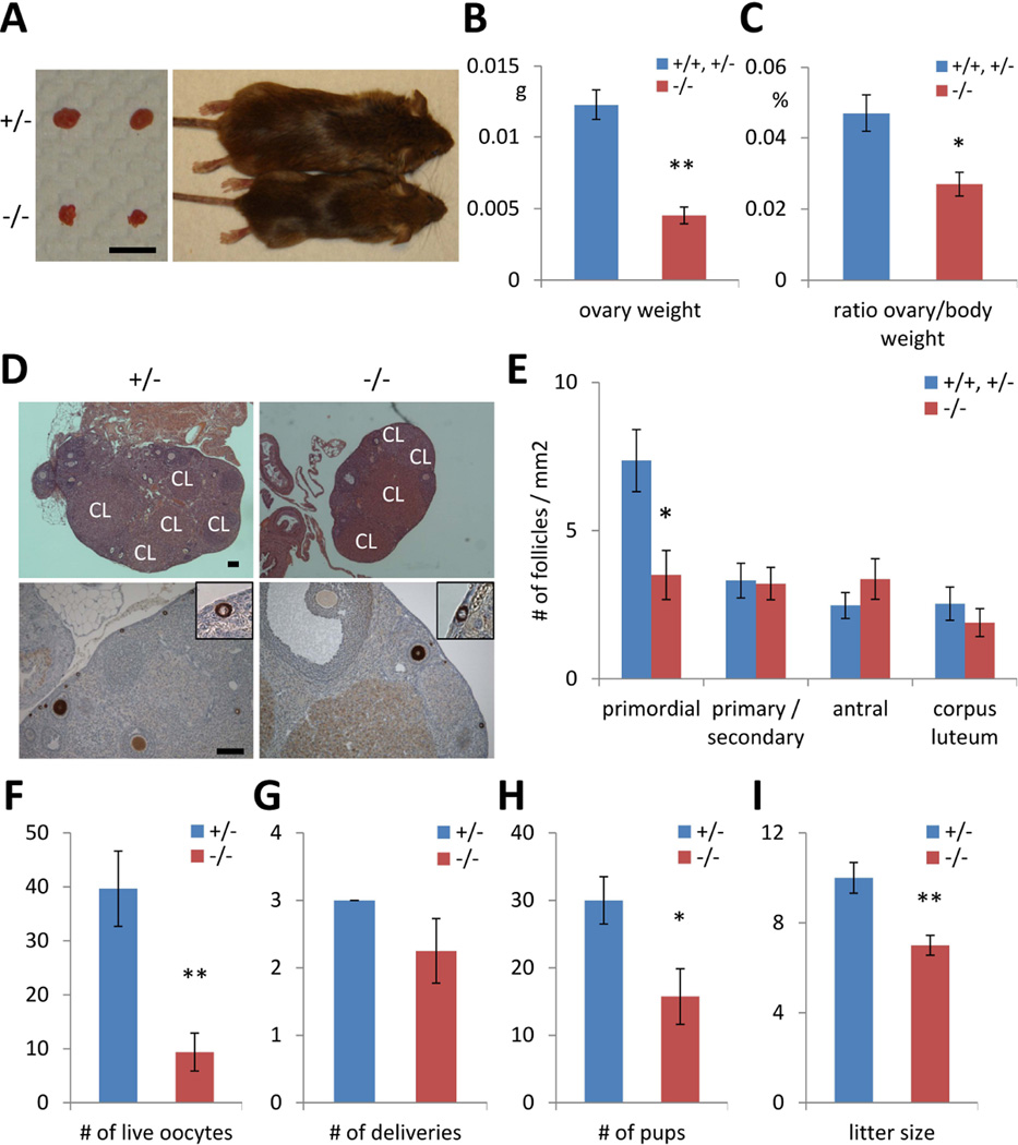
Lin28a knockout females have reduced fertility. (A) A representative photographs of 5-month-old Lin28a +/− and −/− mice and their ovaries. Bar 5mm. (B–C) Ovary weight (B), ovary weight relative to body weight (C) in 5-month-old Lin28a +/− and −/− females. N=4–6. (D) H&E staining (top) and immunostaining with Mvh (bottom) of 3-month-old adult ovaries. Inset: higher magnification of primordial follicles. Note that corpus luteum (CL) are found in both Lin28a +/− and −/− ovaries. Scale bars 100 µm. (E) Number of different stages of ovarian follicles per 1 mm2 in 3-month-old Lin28a +/+, +/− and −/− ovaries. N=8–10. (F) Number of live oocyte collected from 2-month-old Lin28a +/− and −/− females after superovulation. N=5–6. (G–I) Female fertility tests at 2–3 months old. Number of deliveries (G) and number of pups born (H) per Lin28a +/− and −/− female during 10-week experimental period. (I) Litter size of Lin28a +/− and −/− females. N=3–4. * p<0.05, ** p<0.01. Error bars represent SEM.

