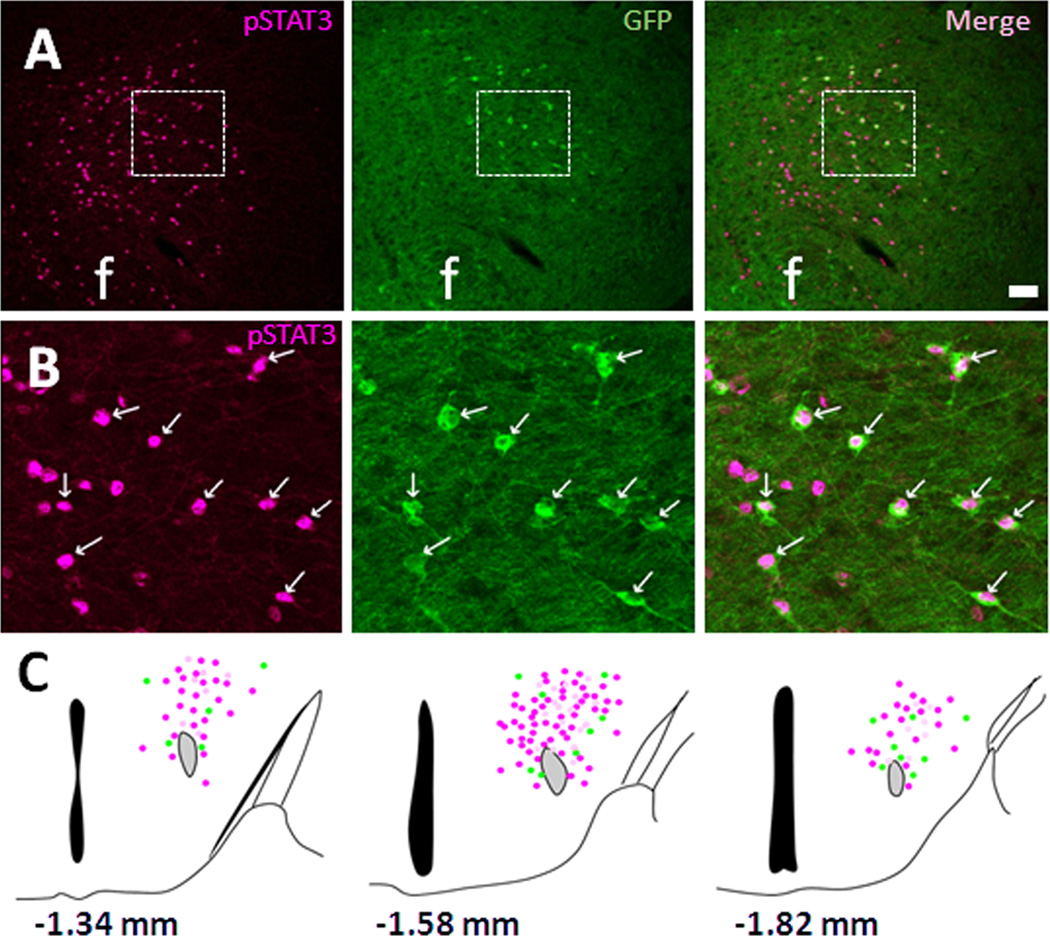
Figure 2. Co-localization of MC4R-GFP neurons with leptin-induced pSTAT3.
Double fluorescent IHC in ICV leptin-treated MC4R-GFP mouse brain sections revealed that leptin-induced pSTAT3 (magenta) and MC4R-GFP (green) are co-expressed in the mid-dorsal LHA (A). Digital zoom of boxed region in (A) is showed in (B). (C) Schematic drawing of distribution of pSTAT3 (magenta), MC4R-GFP (green) and double-positive (white) cells in 3 different levels of the LHA. Note that actual expression of NT and MC4R-GFP cells in other hypothalamic nuclei is not presented here. White arrows indicate representative co-localization. f, fornix; Scale bars: A, 80 um; B, 40 um.

