
Figure 1.
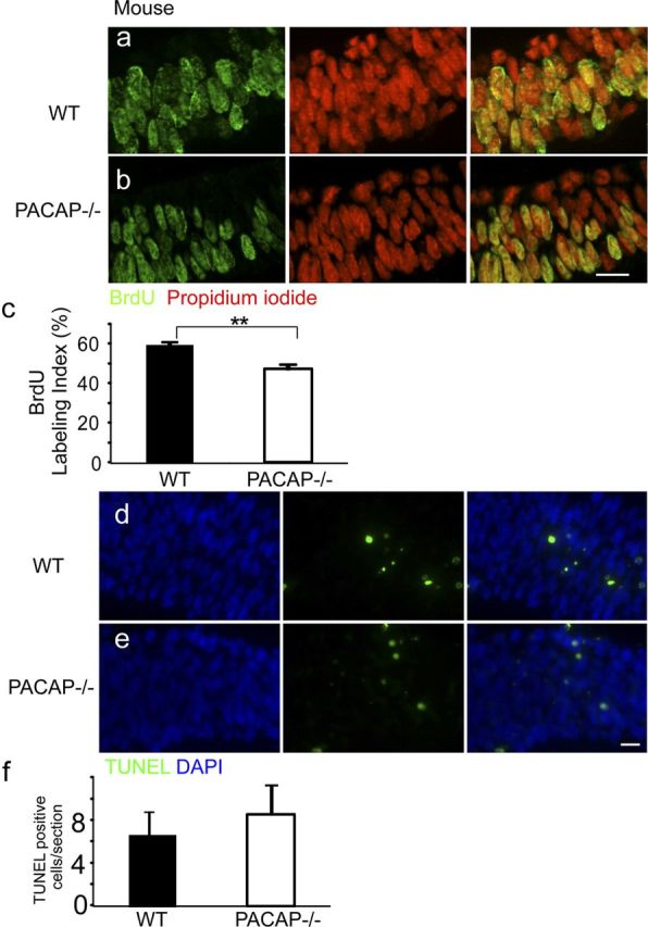
The telencephalic vesicles of E9.5 PACAP−/− mice exhibit reduced S-phase labeling but no change in cell survival. a, b, BrdU immunohistochemical staining of E9.5 WT (a) and PACAP−/− (b) VZ analyzed on coronal sections from the mid-dorsolateral cortex. BrdU-positive cells (green) and total cells (PI; red) were counted blind in the VZ on these confocal images. c, Quantification of BrdU LI. PACAP−/− exhibited a 47% LI compared with 60% in the WT. d, e, TUNEL staining of E9.5 WT (d) and PACAP−/− (e) VZ. TUNEL-positive cells (green) and total cells (DAPI; blue) were counted blind. f, Quantification of TUNEL-labeled cells per section. n = 3 for each genotype. Results are expressed as mean ± SEM. **p < 0.01. Scale bars, 10 μm.