
Figure 10.
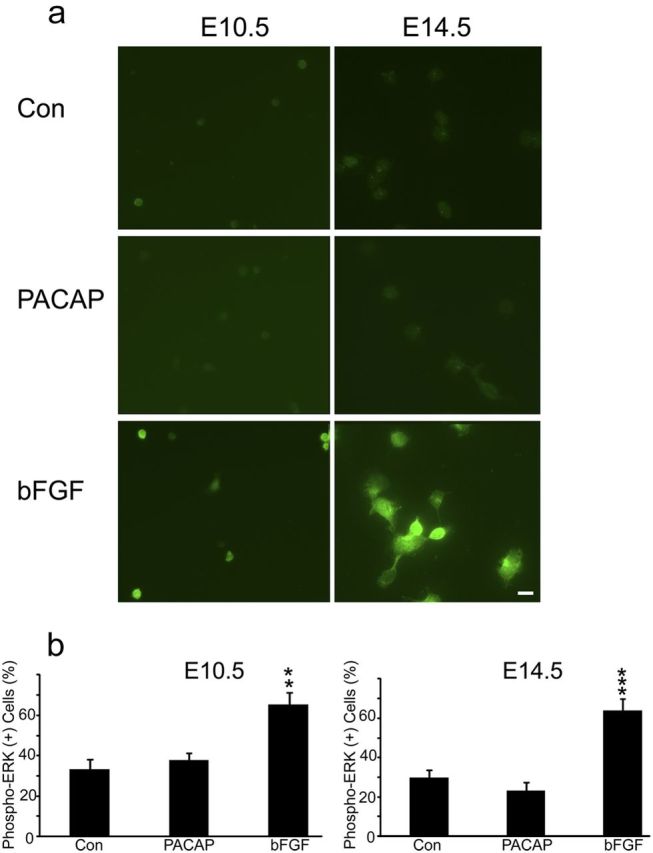
PACAP did not activate phospho-ERK, whereas bFGF exposure led to ERK activation in both E10.5 and E14.5 precursors. a, Examples of phospho-ERK staining in E10.5 and E14.5 10 min after treatment with vehicle (Con), PACAP (10 nm), and bFGF (10 ng/ml). b, Quantification of phospho-ERK immunostaining. Cells were plated in 35 mm dishes in defined media without growth factors, and reagents were added at 2 h for 10 min. Data are representative of three experiments, three dishes per group.**p < 0.01, ***p < 0.001. Scale bar, 10 μm.