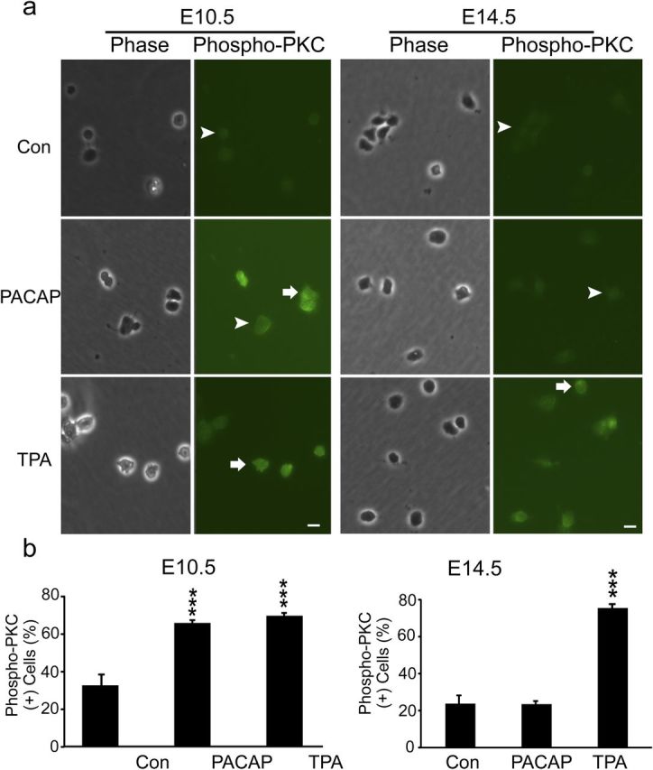
Figure 9.

PACAP activates PKC in early E10.5 cortical precursors but not in E14.5 cells. a, Patterns of phospho-PKC staining in E10.5 and E14.5 cortical precursors 30 min after treatment with vehicle (Con), PACAP (10 nm), and PKC agonist TPA (200 nm). PACAP exposure increased phospho-PKC-positive cells by twofold compared with vehicle in 2 h cultures, comparable with the PKC agonist at E10.5. In contrast, PACAP did not elicit changes in phospho-PKC-positive cells at E14.5, whereas both ages responded to agonist TPA. Positive cells are indicated by arrows and negative cells by arrowheads. b, Quantification of phospho-PKC immunostaining. Cells were plated in 35 mm dishes in defined media without growth factors, and reagents were added at 2 h for 30 min. Data are representative of three experiments, three dishes per group per experiment. ***p < 0.001.
