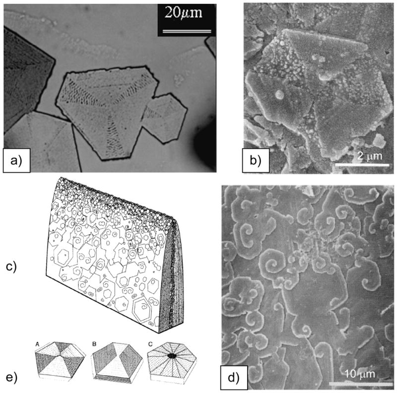
Figure 36.

Comparison of PILP tablets to seminacre tablets. (a) These PILP formed calcite tablets nucleated off the (001) basal plane, and therefore exhibit a hexagonal morphology. Linear transition bars are seen in the wide sectors, while wavy transition bars are seen in the alternate narrower sectors. Some surrounding amorphous film can also be seen above the tablets, which appears to be dissolving in lieu of the crystal tablet. (b) Etching patterns are seen in cyclostome bryozoan seminacre (composed of calcite), where the narrower sectors are preferentially etched. Note the granularity in the etched regions, and finely laminated appearance in the wide sectors. Bar is 2 μm. (c) Schematic illustration by Weedon and Taylor,314 showing the disorganized layers of hexagonal tablets in seminacre, which grow by accretion on all sides, where ‘screw dislocations’ are the dominant mechanism for wall thickening in the older parts of the skeleton. (d) SEM example of the typical surface of seminacre, as described in c). (e) Schematic illustration of sectorization patterns: seminacre tablet divided into six roughly equal, alternately etched sectors (left); nacre tablet divided into a pair of sub-triangular, less-soluble sectors that meet at the center and two rhombic (or paired triangular), more soluble sectors (middle); and gastropod or cephalopod nacre tablet divided into polysynthetically twinned sectors (crystal individuals). The central hollow represents the site of the central organic accumulation (right). (a) (Reprinted with permission from ref 203. Copyright 2000 Elsevier Science B.V..) (b–e) Reprinted from ref 314 with permission from the Marine Biological Laboratory, Woods Hole, MA..)
