Figure 49.

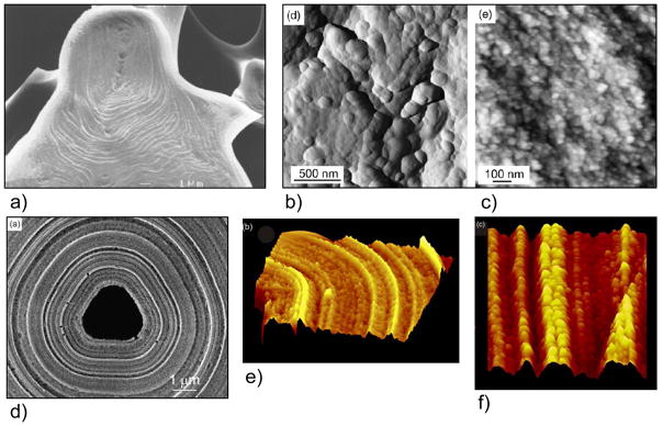
Examples of the concentrically-laminated textures seen in some large biominerals, such as the urchin spine and sponge spicules shown here, and their nanogranular subtextures. (a) SEM of an EDTA-etched fracture surface of an immature sea urchin spine showing the concentric mineral deposition lines. Bar = 1 μm. (b) AFM deflection image showing a nanogranular texture on a naturally grown trabecular surface of the urchin spine. (c) The nanogranular subtexture is also seen in this fracture surface, which may be relevant to the unusual conchoidal fracture patterns seen in urchin spines. (AFM height image, height range ~ 50 nm). (d) SEM of a siliceous sponge spicule. Etching with sodium hypochlorite reveals the concentric laminations of deposited silica, and also removes the organic axial filament that runs down the middle of the spicules. (scale bar 1 μm). (e) AFM surface plots of a spicule cross-section reveals the nanogranular subtexture within the concentric layers. (scan size 3.8 μm, height data scale 275 nm). (f) AFM surface plot of a longitudinal section revealing details of the annular nanoparticulate substructure (scan size 2 μm, height data scale 75 nm). (a) (Reprinted with permission from ref 54. Copyright 1997 The Royal Society of Chemistry.) (b–c) (Reprinted with permission from ref 345. Copyright 2008 Elsevier Ltd..). (d–f) (Reprinted with permission from ref 346. Copyright 2003 Elsevier Inc..)
