
Figure 53.
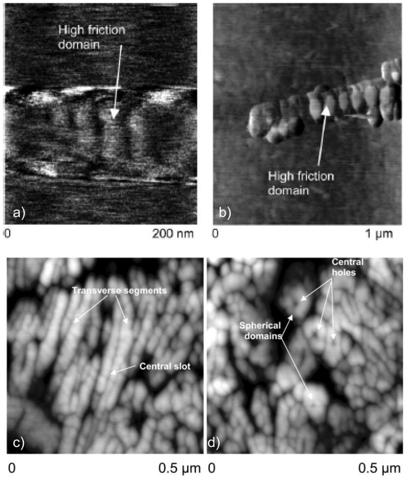
Nanoscale textures in dental enamel crystallites, as observed by Robinson et al.380. (a & b) Frictional force imaging of maturation stage rat enamel crystals. (a) Friction obtained using hydroxylated cantilever tips, pH 7. Lighter areas, which correspond to regions of high friction, show transverse high friction bands ~40 nm in width. (b) At at pH 5.5, the banding is again visible, but some bands exhibited patches of much higher friction. (c & d) High resolution AFM height images of polished human enamel sections. Images were obtained with unmodified tips, tapping mode in air. (c) Longitudinal/oblique sections of enamel crystal. Some transverse segmentation of crystals is visible indicative of the banding structure seen on maturation stage crystals. Central dark lines represent holes or grooves along the crystal long axes. (d) Transverse sections of human enamel crystals. The crystals appear to be made up of a series of roughly spherical subunits surrounding a central hole or pit. (Reprinted with permission from ref 380. Copyright 2004 The Royal Society of Chemistry.)