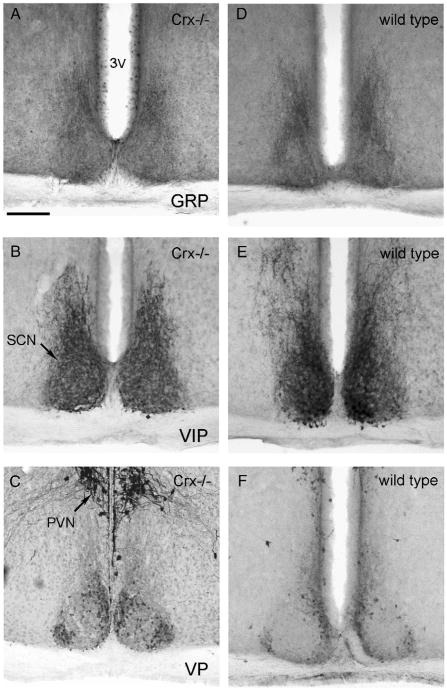
Fig. 2.
Photomicrographs of the suprachiasmatic nuclei of the Crx−/− (left column) and wild type (right column) mouse. The sections have been immunohistochemically reacted for gastrin releasing peptide (GRP, A and D), vasoactive instestinal peptide (VIP, B and E), and vasopressin (VP, Figs. 2C and F). 3V=third ventricle, PVN=paraventricular nucleus, SCN=suprachiasmatic nucleus. Bar=200 μm.

