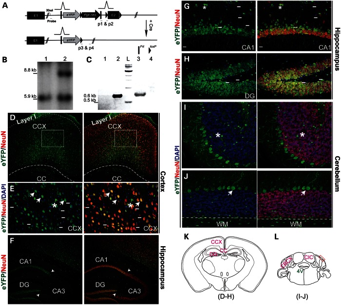
Figure 2.
Expression of Pk1 in adult brain, revealed by an eYFP reporter driven by the endogenous promoter. (A) An eYFP reporter gene, PGK-neo cassette, Frt and loxP sites are targeted into the Pk1 exon 2 region in the order shown here. An En1 gene splicing acceptor is placed before eYFP start codon and several polyadenylation signals are after eYFP stop codon. This gene-trap allele can be excised by Cre to generate a straight knockout allele. The Sox2-Cre transgenic line is used to cross with Pk1 gene-trap mice in this study. (B) Southern analysis of XhoI-digested genomic DNA using a 5′ probe reveals an 8.8 kb band, showing correct integration of the knock-in gene-trap construct into the Pk1 locus (lane 2) and a 5.9 kb fragment for the WT allele (lane 1). (C) PCR genotyping of gene-trap and straight knockout alleles after Cre excision. Primers 1 and 2 will amplify a 0.6 kb fragment from the straight KO allele (lane 2). Primers 3 and 4 will amplify a 0.65 kb fragment from the gene-trap allele (lane 3). L, DNA ladder. Lanes 1 and 4 are wild-type controls with no PCR products. (D–J) Images shown in these panels are from adult brain at 4-week-old. (D) Pk1 is broadly expressed in the cerebral cortex, as identified by eYFP-positive cells, except for layer I. A majority of the eYFP-positive cells (green) are NeuN positive (red). Boxed areas are shown in (E). (E) High magnification shows expression of eYFP in both neurons (NeuN positive, arrows) and glia (asterisks). Short lines indicate a fraction of eYFP-negative neurons. Note that most eYFP-positive neuronal somata are triangular, a typical shape for pyramidal neurons. (F) Pk1 expression in hippocampus, with strong labeling in CA1 and DG and weaker expression in CA3 (arrowheads demarcate the boundary). (G and H) A high magnification image shows overlapping of Pk1 expression (green) with NeuN in CA1 and DG. Note that some NeuN-positive neurons in the molecular layer are eYFP negative (above the short lines). (I and J) Cerebellar Pk1 expression in Purkinje, but not in granular neurons. Purkinje neurons are NeuN negative; granular neurons are NeuN positive (red). Asterisks indicate glia; arrows indicate Purkinje cells. Below the dashed lines, we observe WM showing sparsely distributed glia (green dots). (K and L) Schematic illustrations of sections of imaged areas for the relevant panels, as indicated below the drawings. 4V, 4th ventricle; red box is the photo area in F and G; CIC, central nucleus of inferior colliculus; CBCX, cerebellar cortex.

