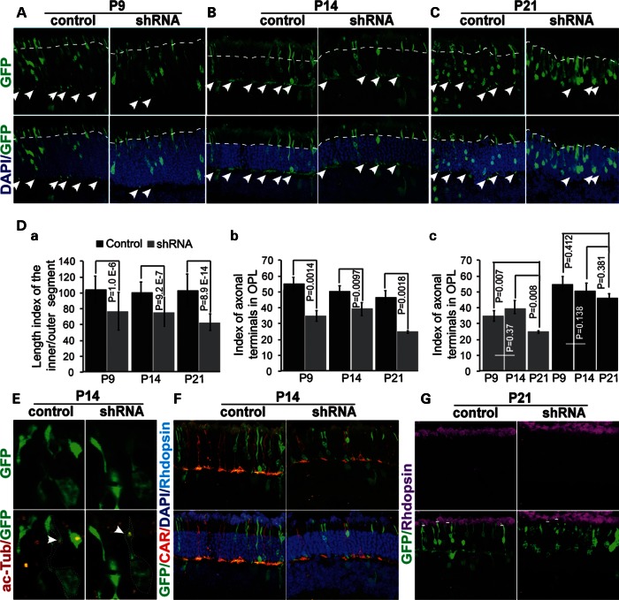
Figure 7.
Rod photoreceptor defects in Pk1 shRNA electroporated retina in vivo. (A). Shortened inner/outer segments (above dashed lines) and a reduced number of axon terminals at P9 caused by Pk1 shRNA (arrows in the inner plexiform layer, IPL). (B) Similar defects are observed at P14 compared with P9. (C). Severe defects in both inner/outer segments and synaptic terminals at P21 (dashed lines and arrows). (D) (a) A significant decrease of inner/outer segments’ length in shRNA electroporated retina at different ages. A length index is generated by measuring the inner/outer segments of GFP-positive photoreceptors using ImageJ. (b) A significant decrease of synaptic terminals in shRNA-electroporated retina at different ages. The axon/synaptic terminal index is the ratio of GFP dots in the OPL to the total GFP-positive cells on the section, multiplied by a factor of “100”. (c) Progressed reduction of axonal terminals in shRNA electroporated retinas occurs between P14 and P21, but not P9 and P14. The original data used to plot the graph are the same as in (b). (E) Acetylated alpha tubulin (red) stains cilia in both control and Pk1-shRNA photoreceptors (green). (F) Pk1-shRNA rods (green) do not change to cones (red) identified by cone arrestin. (G) Pk1-shRNA photoreceptors still express rhodopsin (above the short lines).

