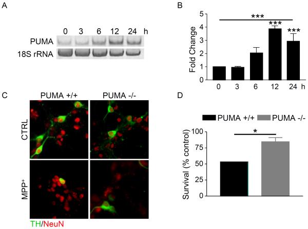
Figure 1. Loss of PUMA attenuates MPP+-mediated cell death.
A) Cultures derived from C57Bl/6 mice were treated with 1 μM MPP+ and total RNA was collected at the indicated times. Levels of PUMA and 18S rRNA were analyzed by semi-quantitative RT-PCR. All reactions were performed in triplicate and three independent experiments were performed. A representative gel is shown. B) Gels were quantitated in ImageQuant and analyzed by one-way ANOVA (***, p < 0.001) with Bonferroni post-tests to compare each time point to untreated (12 hr and 24 hr: ***, p < 0.001). C) Cultures derived from PUMA +/+ and PUMA −/− cultures were treated with 1 μM MPP+ for 48 hours. Cells were fixed and stained for TH and NeuN. D) TH-positive and NeuN-positive cells were counted, the percentage of TH-positive cells was calculated and survival expressed as a percentage of untreated control. Data were analyzed by Student's t-test (*, p < 0.05).

