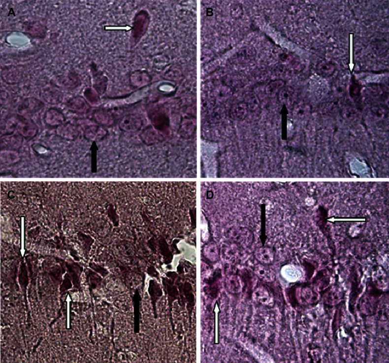
Fig 3.
Photomicrographs of coronal sections of the CA1 region of the hippocampus (caspase-3 immunohistochemistry, counterstain with nuclear fast red). A: sham group; B: sham+CA group; C: lesion group; and D:lesion+CA group. Black arrows show intact pyramidal cells and white arrows show apoptotic pyramidal cells that express positive to caspase-3 antibody ×1000.

