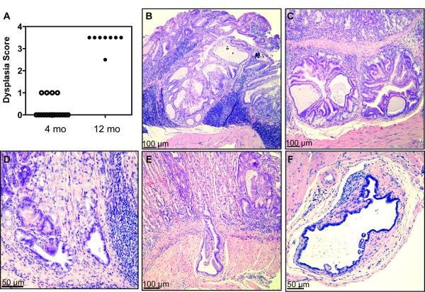
Figure 6. TxA23 mice develop masses with dysplastic foci as they age.
(A) Dysplasia scores (where 1 = irregular glad forms, 3 = severe loss of glands and of columnar orientation of epithelium with regions of cellular atypia, increased mitotic figures, and 0.5 added when invasion of muscle or carcinoma in situ is identified) for sections at give ages. Each point represents an individual mouse. (B) Section of a mouse at 10 months of age (magnified 10×). Note to formation of mass with abundant, dense chronic inflammatory infiltrates. (C-F) Images of stomach sections that represent pathology observed in 12 month old mice with various degrees of pseudoinvasion of irregular glands into the submucosa. (C) Section showing submucosal focus of irregular gland formations (magnified 10×). (D) Section showing focus of irregular glands in submucosal tissue with surrounding chronic inflammation (magnified 20×). (E) Section showing deep submucosal pseudoinvasion by an irregular gland (magnified 10×). (F) Irregular glandular form on the adventitial surface of the stomach (magnified 20×).

