
Figure 1.
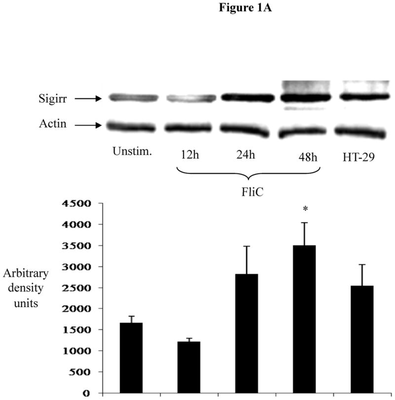
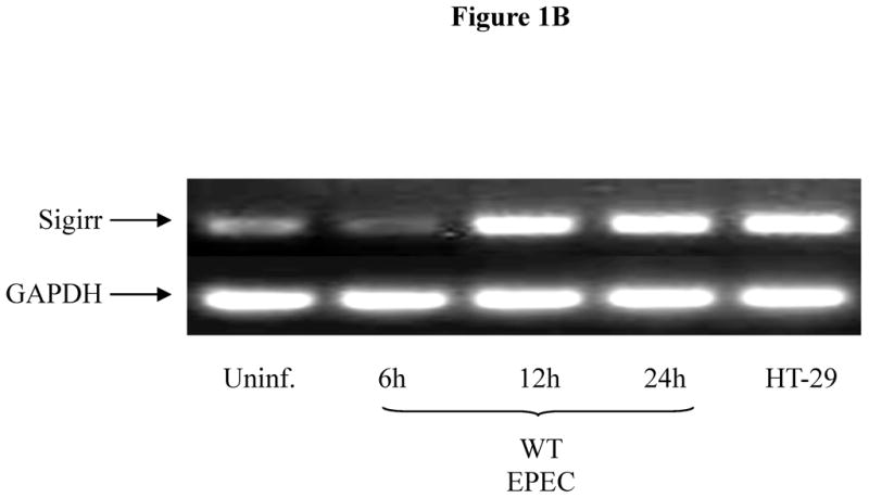

Figure 1A: Exposure to bacterial flagellin induces Sigirr protein expression in non-transformed human colonic (NCM) IEC. NCM were grown in M3 growth media and exposed to 10 ng/ml purified flagellin (FliC). Cell lysates were prepared as described in materials and methods. 30–50μg of cleared cell lysates were subjected to SDS-PAGE and probed with rabbit polyclonal antibody to Sigirr. Blots were developed in ECL and bands analyzed by densitometry. Result representative of three separate experiments.
Figure 1B: EPEC infection increases Sigirr message levels in Caco-2 IEC. Cells were grown to confluence in 12 WP and infected with WT EPEC for 3 h. After infection, cells were washed twice and fresh media added with gentamycin to prevent bacterial growth. Total RNA extracted at different time-points was subjected to semi-quantitative RT-PCR to measure gene expression as described in Materials and Methods. GAPDH was included as internal control. Blot representative of three independent experiments.
Figure 1C: EPEC infection increases IL-8 expression in Caco-2 IEC. Cells were grown to confluence in 12 WP and infected with WT EPEC for 3 h as above. Afterwards, cells were washed twice and fresh media added with gentamycin. Total RNA was extracted and subjected to semi-quantitative IL-8 RT-PCR. GAPDH was included as internal control. Blot representative of two separate experiments.