
Figure 2.


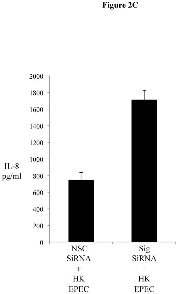
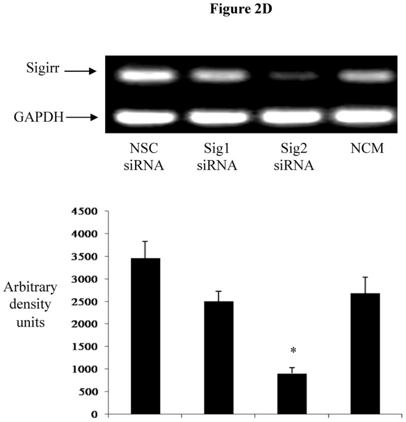


Figure 2A: Transient gene silencing reduces Sigirr protein in Caco-2 cells. IEC (2 X 10⁵/well) were seeded in 24 WP. After 48h, cells were transfected with Sigirr and non-silencing control (NSC) siRNA in Hiperfect transfection reagent. Sigirr protein was analyzed by SDS-PAGE as described in materials and methods and developed in ECL. Bands were quantified by densitometry with BioRad quantity one software program.
Figure 2B: Sigirr gene silencing augments flagellin induced IL-8 chemokine secretion from Caco-2 cells. Sigirr and NSC siRNA transfected Caco-2 cells were exposed to 10 ng/ml purified flagellin (FliC) and Interferon gamma (IFNg) (100 ng/ml) in DMEM with serum and antibiotics. IL-1β (5 ng/ml) was included as a positive control. Cell culture supernatant was collected after 6h and IL-8 quantified by ELISA. Error bars and * p<0.05 vs. NSC siRNA + FliC.
Figure 2C: Heat-killed (HK)-EPEC-induced IL-8 production is enhanced upon Sigirr gene silencing in Caco2 cells. . Sigirr and NSC siRNA transfected Caco-2 cells were exposed to HK-EPEC in DMEM with serum and antibiotics. Cell culture supernatant was collected after Xhr and IL-8 quantified by ELISA. Error bars = ?.
Figure 2C 2D: Transient gene Silencing decreases Sigirr gene expression in HT-29 cells. IEC (2 X 10⁵/well) were seeded in 24 WP and transfected after 48 h with Sigirr and non-silencing control (NSC) siRNA. Total RNA was extracted and Sigirr gene expression measured by semi-quantitative RT-PCR. GAPDH was included as internal control. Blot is representative of three experiments and bands were analyzed by densitometry
Figure 2D 2E: Sigirr gene silencing augments TLR5 mediated IL-8 secretion. siRNA transfected HT-29 cells were exposed to FliC (10 ng/ml), in presence and absence of a specific TLR5 neutralizing antibody. Cell culture supernatant was collected after 6 h and IL-8 quantified by ELISA. IL-1β (5 ng/ml) was included as positive control. Error bars and * p<0.05 vs. NSC siRNA + FliC and Sig siRNA + FliC.
Figure 2E 2F: Sigirr gene silencing augments diverse TLR responses in IEC. siRNA transfected HT-29 cells were exposed to FliC (10 ng/ml), LPS (20 ng/ml), Pam3Cys (Pam-20 ug/ml) and Phorbol myristate acetate (PMA-50 ng/ml) for 6 hours in DMEM with serum and antibiotics. Cell culture supernatant was collected and IL-8 quantified by ELISA. Error bars and * p<0.05 vs. NSC siRNA + FliC, NSC siRNA + LPS and NSC siRNA + Pam.
Figure 2F 2G: EPEC infection up-regulates chemokine response in Sigirr deficient HT-29 cells. IEC were grown to confluence and infected with wild type EPEC for 3 h. After infection, cells were washed twice and fresh media added with gentamycin treatment. Cell culture supernatant was collected after 12 h for IL-8 ELISA. Error bars and * p<0.05 vs. NSC siRNA + EPEC.