
Figure 5.

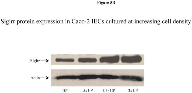
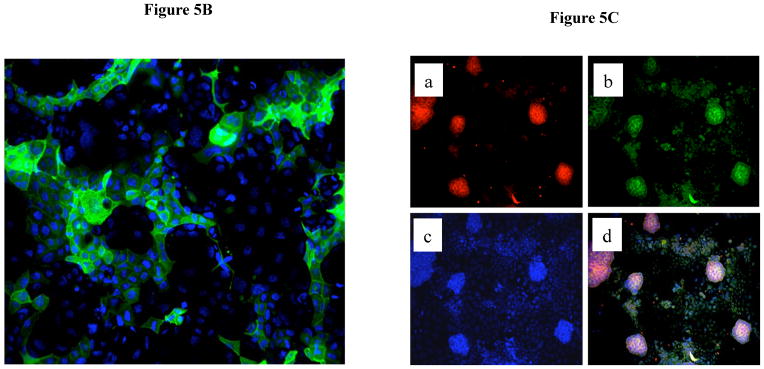
Sigirr expression in Caco-2 cells is dependent on the differentiation state. (A) Caco-2 monolayers were exposed to sodium butyrate (2mM) for 24–72 h in DMEM supplemented with serum and antibiotics. 30–50μg of total protein from pre-confluent, sodium butyrate treated and spontaneously differentiating post-confluent cells were probed with Sigirr rabbit polyclonal antibody in western blots. Result representative of two experiments. (B) Caco-2 cells were seeded at increasing densities as indicated, and xug of total protein from post-confluent cells were probed with Sigirr antibody in western blots as above. (C) Caco-2 cells express the differentiation marker Dipeptidyl dipeptidase (DPP) in IEC monolayers. Formalin fixed Caco-2 monolayers grown on coverslips were incubated with mouse monoclonal DPP antibody conjugated with Alexa fluor-488 (green). Cell nuclei stained with dapi. Magnification 40X. Result representative of three independent experiments. (D) Sigirr co-localizes with the differentiation marker DPP in Caco-2 cells. Cells grown higher density (5 × 10⁵/well) were fixed in 4% formalin and incubated simultaneously with goat polyclonal Sigirr antibody (a-red) with mouse monoclonal DPP antibody conjugated to Alexa fluor 488 (b-green) and nuclear dapi stain (c-blue). Images taken with Zeiss fluorescent microscope were merged to show co-localization (d). Result representative of two independent experiments. Magnification 20X.