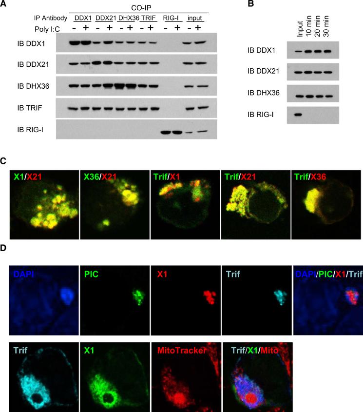
Figure 4. Identification of the DDX1-DDX21-DHX36-TRIF Complex in Cells.
(A) Whole-cell lysates from D2SC cells, with or without poly I:C stimulation, were incubated with the indicated antibodies and protein G beads. Bound proteins were analyzed by immunoblotting with the indicated antibodies.
(B) D2SC cells were incubated with bio-poly I:C for 10, 20, or 30 min. Whole-cell lysates from the treated D2SC cells were prepared and subjected to purification with NA-beads. The proteins bound to bio-poly I:C were detected with indicated antibodies.
(C) HEK293T cells were cotransfected with indicated expression vectors. Cells were stained with Myc or HA antibodies.
(D) HEK293T cells were cotransfected with indicated expression vectors. Twenty-four hours later, cells were stimulated with poly I:C or Alexafluor 488 labeled poly I:C for 4 hr, then stained with Myc antibody or HA antibody. MitoTracker was used to probe the mitochondrion. DAP1 served as the nuclei marker.

