
Figure 4.
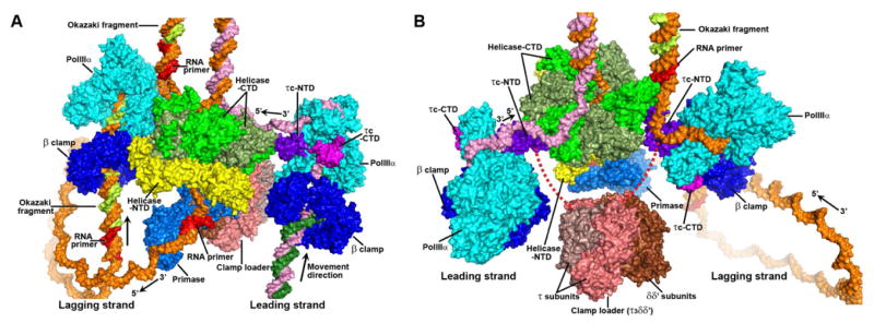
An atomic model of the replisome structure at the replication fork. (A and B) The surface representations of these replisome components are displayed along two different directions. PolIIIα, β clamp, the NTD of τc, the CTD of τc, primase, and the NTD of helicase are labeled with cyan, blue, purple, magenta, marine and yellow respectively. The CTDs of helicase are labeled with green and darkgreen. The five subunits of clamp loader are labeled with salmon, deepsalmon, and brown. The red dash lines represent the loop linking the two domains of the τ subunit. The modeled DNA strands are labeled with orange and pink for the mother strands, and with lime and forest for the daughter ones. The RNA primers are labeled with red. See also Movie S1.