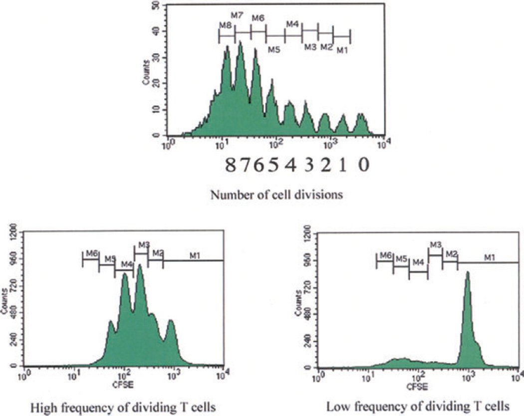
Fig. 3.
Flow cytometry analysis of cell proliferation by CFSE labeling: Following lymphocyte activation and proliferation, each cell division results in a halving of the fluorescence intensity of the intracellular fluorescent dye CFSE. This sequential halving of cellular fluorescence intensity is visualized as distinct peaks or populations of cells and can be used to track cell division in populations of proliferating cells. In T lymphocytes, up to eight cell divisions can be accurately distinguished. CFSE, carboxyfluorescein succinimidyl ester.

