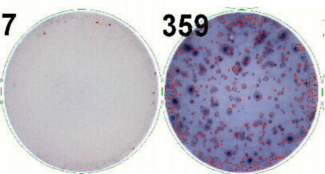
Fig. 4.
ELISPOT assay: Cells are incubated in plates coated by a capture antibody specific for the cytokine of interest. Following a period of incubation, the cells are washed away and the cytokine remains bound to the antibody. The bound cytokine is detected using labeled secondary antibodies. In the final step, a substrate is added that precipitates where the secondary antibody was bound, forming spots that correspond to cells producing the cytokine.

