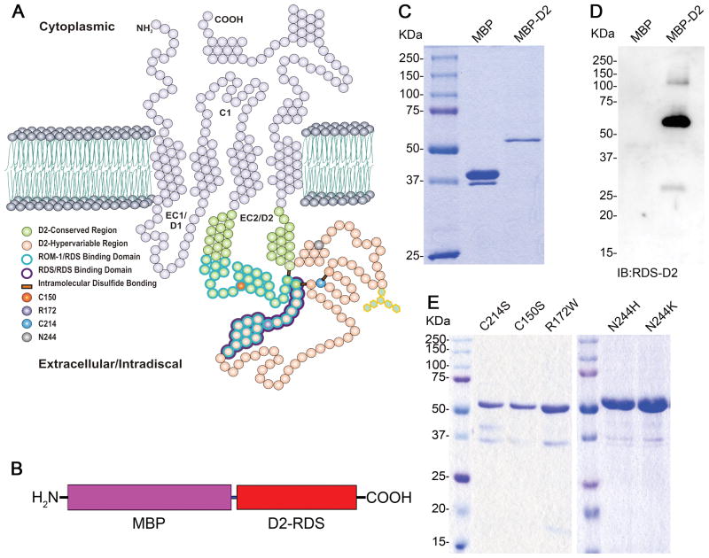
Figure 1. Generation and purification of RDS D2 loop fusion proteins.
(A) Structural features of RDS protein. RDS contains four transmembrane domains, two intradiscal loops (D1 and D2), cytoplasmic N- and C- termini, and a small cytoplasmic loop (C1). (B) Schematic representation of MBP-fusion protein. The full-length D2 loop of RDS consisting of residues Phe120-Asn256 corresponds to the red bar labeled D2-RDS. Purified MBP (middle lane) and MBP-D2 (right lane) were subjected to (C) SDS-PAGE and stained with Coomassie brilliant blue, and (D) western blot analysis using polyclonal anti-D2 (RDS) antibody. (E) Different mutants of RDS D2 (C214S, C150S, R172W, N244H and N244K) were expressed and purified as MBP fusion proteins. Purified proteins were subjected to SDS-PAGE and stained with Coomassie brilliant blue to check the purity. The mutant proteins were also highly expressed in this system and migrated to the same position as MBP-D2 -WT in the gel.

