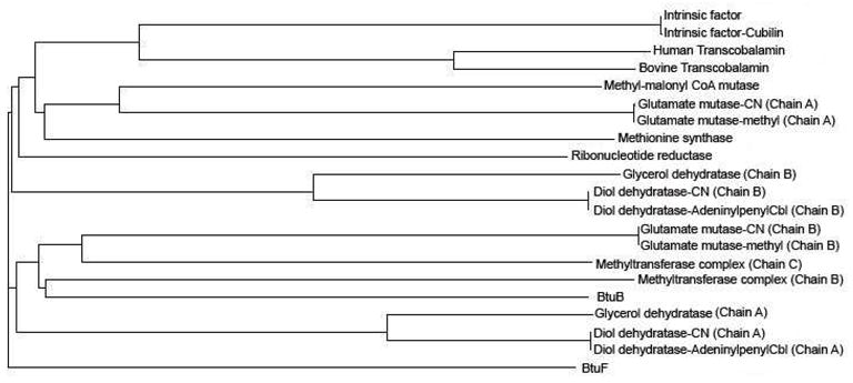
Figure 8.

(a) Worms/tubes diagram of Methylmalonyl-CoA mutase. The α-chain and β-chain areshown in gold and green color respectively. B12 is shown as ball and stick in black color (b)Worms/tubes diagram of Glutamate mutase. The σ subunit is shown in red and green colors,while the ε subunit is shown in gold and cyan colors. B12 is shown as ball and stick inblack color (c) Worms/tubes diagram of Diol dehydrate. B12 is shown as ball and stick in blackcolor. (d) Worms/tubes diagram of ribonucleotide reductase. B12 is shown as ball and stick in blackcolor.
