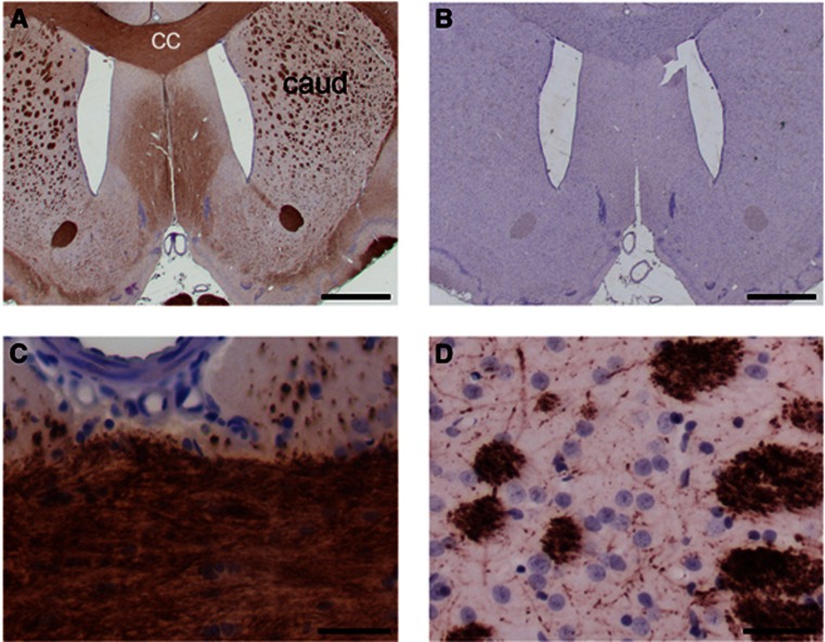
Figure 2.
Histological examination of white matter. Immunohistochemical labeling of myelin basic protein (MBP) in brain sections from a representative stroke-prone spontaneously hypertensive rat (SHRSP), aged 47 weeks. (A) MBP immunolabeling (brown) displays histological white matter areas in low magnification view of a coronal section. The corpus callosum (cc) and caudate (caud) are marked. (B) A neighboring section treated identically but without primary antibody shows no immunoreactivity. (C, D) Higher magnification images confirm integrity of myelin in corpus callosal white matter (C) and in caudate white matter bundles (D). The central azimuthal artery serves as an anatomical landmark in (C). Nuclear chromatin is counterstained with hematoxylin (blue). Scale bars: 1,000 μm (A, B) and 20 μm (C, D).

