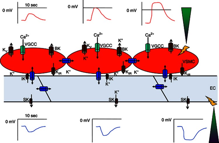
Figure 2.
Conduction mechanism involved in vascular conducted responses (VCRs). The schematic shows coupled endothelial cells (ECs) and vascular smooth muscle cells (VSMCs) in the arteriolar wall, equipped with ion channels and gap junctions necessary to sustain conduction through the intercellular compartment(s) along the vessel. On the right, two pipette tips (green color) show delivery of a depolarizing (upper panel, VSMC) and hyperpolarizing (lower panel, EC) current to initiate conduction along the vessel. Both the depolarization (red curve) and hyperpolarization (blue curve) is seen to decay with distance from local stimulus. Blue barrels depict gap junctions coupling EC–EC, and VSMC–VSMC, as well as myoendothelial coupling (EC–VSMC). Green barrels depict voltage-gated Ca2+ channels (VGCC) whose activity convert the de- or hyperpolarizations into an increased or decreased Ca2+ influx into VSMC. Brown barrels depict various K+ channels in EC and VSMC, whose function is to modify the electrical responses as they are conducted along the EC and VSMC pathways. See text for further explanations. BK, big conductance Ca2+-activated K+ channels; IK, intermediate conductance Ca2+-activated K+ channels; SK, small conductance Ca2+-activated K+ channels; KV, voltage-gated K+ channels; KIR, inward-rectifier K+ channels. The duration bar is arbitrarily set to 10 seconds in the plot of Vm versus distance. The ECs and VSMCs are not drawn to scale.

