
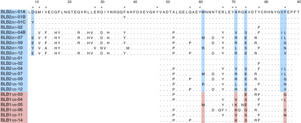
Figure 2.
Alignment of amino acid sequences. PBR positions from Tong et al. [73] are marked with a +. The shaded amino acids are the PAML derived positively selected codon positions, blue for BLB2 and pink for BLB1.
Official websites use .gov
A
.gov website belongs to an official
government organization in the United States.
Secure .gov websites use HTTPS
A lock (
) or https:// means you've safely
connected to the .gov website. Share sensitive
information only on official, secure websites.
Alignment of amino acid sequences. PBR positions from Tong et al. [73] are marked with a +. The shaded amino acids are the PAML derived positively selected codon positions, blue for BLB2 and pink for BLB1.