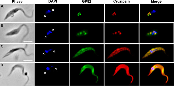
Figure 3.

Colocalization of GP82 with cruzipain during metacyclogenesis. Immunofluorescence showing intermediate forms attached to culture flasks at 48 h (A-B) and late intermediate forms and fully differentiated metacyclic forms in culture supernatant (C-D) at 48 h after nutritional stress. Cells were fixed, permeabilized with 0.5% saponin and submitted to immunofluorescence using mAb 3F6 and rabbit polyclonal antibody against T. cruzi cruzipain. DAPI was used to stain nucleus (N) and kinetoplast (K). Scale bar = 10 μm.
