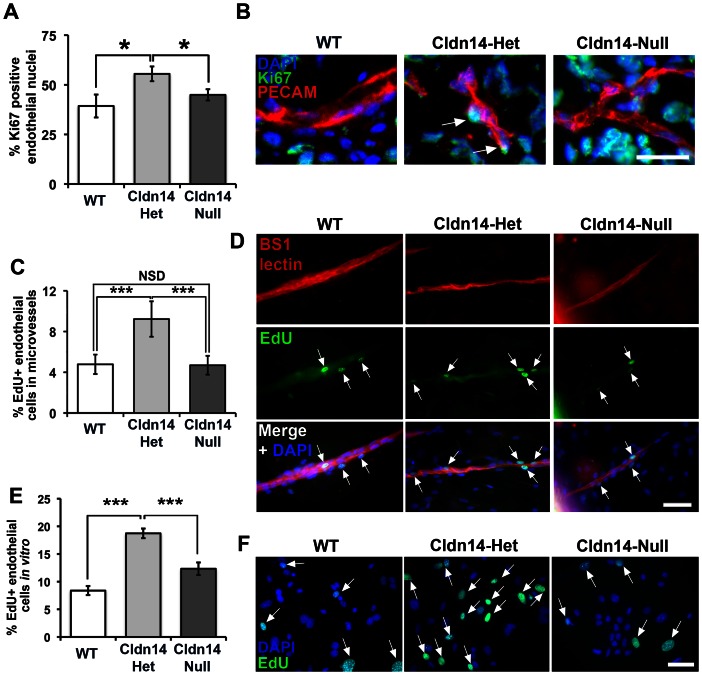
Figure 6. Cldn14 gene copy number affects endothelial cell proliferation in vivo, ex vivo and in vitro.
(A) Percentages of Ki67-positive endothelial cells were counted in cryosections of 13-day B16F10 tumours from WT, Cldn14-het and Cldn14-null mice co-stained with PECAM. Endothelial cell proliferation was enhanced significantly in Cldn14-het mice. (B) Representative images of tumour sections in each genotype. Arrows, Ki67-positive endothelial cell nuclei. Scale bar 25 µm. (C) Proliferating cells in VEGF-stimulated wild-type, Cldn14-het and Cldn14-null collagen-embedded aortic explants were detected by EdU incorporation. The number of proliferating (EdU-positive) nuclei, counterstained with DAPI, was divided by the total number of cell nuclei also BS1-lectin positive to give % proliferating endothelial cells in VEGF-treated aortic rings. Bars show mean % of proliferating cells ± SEM. n = 6–8 rings per genotype, 513–717 nuclei per genotype. (D) Representative images of VEGF-stimulated WT, Cldn14-het and Cldn14-null microvessels from aortic ring explants stained for EdU and BS1 lectin. Scale bar 50 µm. (E) WT, Cldn14-het and Cldn14-null primary endothelial cells were examined for EdU incorporation in the presence of 30 ng/ml VEGF. Cells were counterstained with DAPI and the number of EdU-positive cells recorded for each genotype. Bars show mean % EdU-positive cells ± SEM. N = 1217–3464 nuclei per genotype, 3 mice per genotype. (F) Representative images of primary endothelial cells in culture. Scale bar 50 µm. Arrows, EdU-positive nuclei. NSD: no significant difference. * P<0.05, *** P<0.001.

