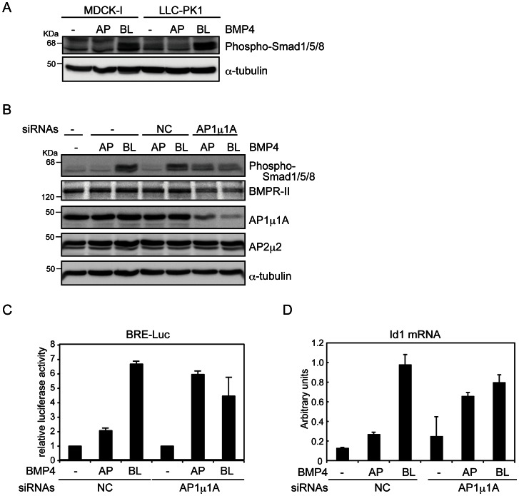
Figure 3. Roles of AP1 µ1A in basolateral trafficking of BMPR-II.
(A and B) MDCK-I cells and LLC-PK1 cells transfected with either control (NC) or AP1 µ1A siRNAs were grown to confluence on Transwell plates, and then treated with 20 ng/ml BMP4 from the apical (AP) or basolateral (BL) sides for 45 min. Phosphorylation of Smad1/5/8 induced by BMP4 was examined by immunoblot analyses using the indicated antibodies. (C and D) LLC-PK1 cells and LLC-PK1 cells transfected with BMP-responsive element (BRE)-reporter construct in combination with either control siRNA (NC) or AP1 µ1A siRNA were seeded on Transwell plates and grown to confluence. Cells were treated with BMP4 (20 ng/ml) from the apical (AP) or basolateral (BL) sides for 2 h for luciferase assays, normalized to activity of sea pansy luciferase encoded by pRL-TK-Renilla (C), and for 1 h for quantitative RT-PCR analysis, normalized to the amounts of GAPDH mRNA (D). Each value represents the mean ± S.D. of triplicate determinations from a representative experiment. Similar results were obtained in two independent experiments (C and D).

