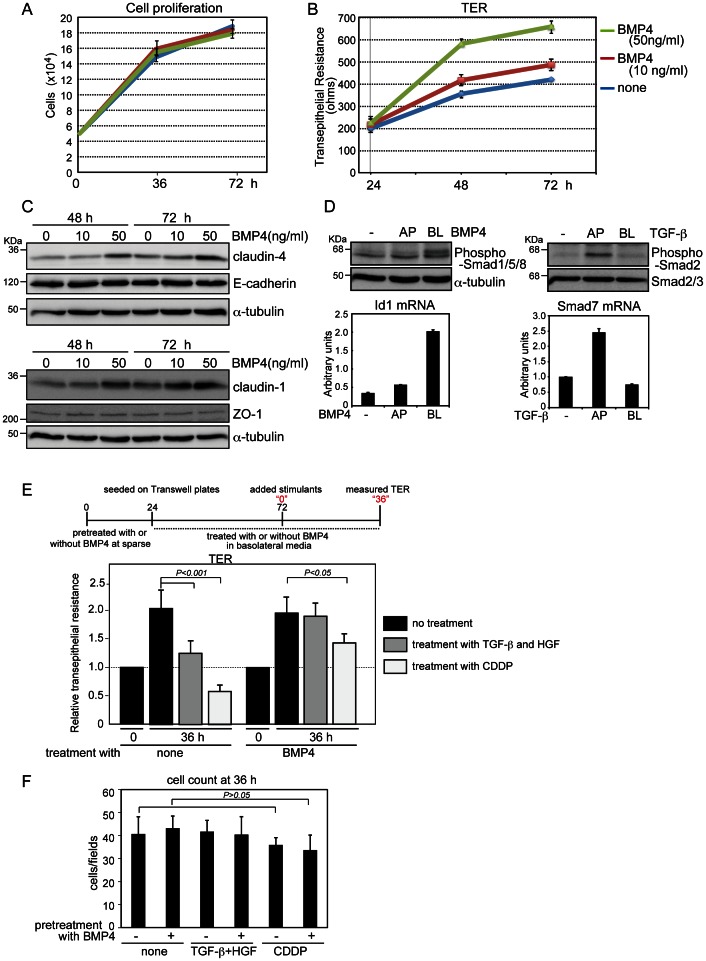
Figure 4. Increase of transepithelial resistance (TER) by BMP4 treatment.
(A, B, and C) MDCK-I cells pretreated with BMP4 for 24 h were seeded on Transwell plates in basolateral media containing BMP4, and incubated until 72 h (i.e., for an additional 48 h). Cell counting (A), TER measurement (B), and immunoblot analyses (C) were performed at the indicated time points. (D) MDCK-I cells were grown to confluence on Transwell plates and treated for 45 min with 20 ng/ml BMP4 (left) and 1 ng/ml TGF-β (right) from the apical (AP) or basolateral (BL) sides. Phosphorylation of Smads and expression of representative target genes (Id1 for BMP4 and Smad7 for TGF-β) were examined by immunoblot and quantitative RT-PCR analyses, respectively. Each value represents the mean ± S.D. of triplicate determinations from a representative experiment. Similar results were obtained in two independent experiments (A, B and D). (E and F) MDCK-I cells pretreated with BMP4 under sparse conditions for 24 h were seeded in triplicate on Transwell plates in basolateral media containing 50 ng/ml BMP4 for 48 h, and TER was measured (indicated as “0”). Then the cells were treated for 36 h either with both 1 ng/ml TGF-β from the apical side and 10 ng/ml HGF from the basolateral side or with 25 µM CDDP from the basolateral side. After TER of all three Transwell plates had been measured at four points for each well (E), the cells from two plates were used for cell counting (F) and the other was used for E-cadherin staining (Fig. S3). Each value represents the mean±S.D. of duplicate determinations from a representative experiment. Similar results were obtained in three independent experiments.

