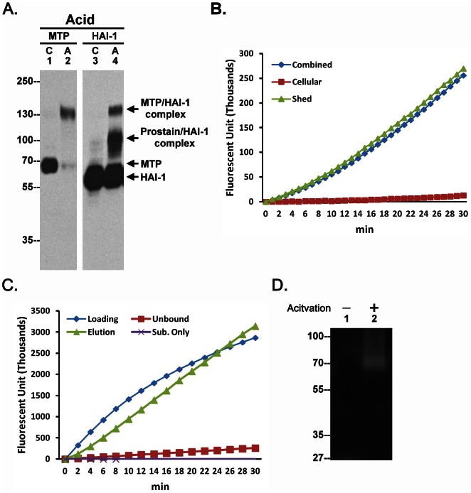
Figure 1. Shedding of active matriptase by human keratinocyte.
A. Human keratinocyte HaCaT cells were incubated either with PBS (C) as a control or a pH 6.0 buffer (Acid, A) to induce activation of matriptase. Cell lysates were analyzed by immunoblot for matriptase (MTP) or HAI-1 (HAI-1). B. HaCaT cells were incubated with a pH 6.0 buffer to induce matriptase activation. The cells and the conditioned buffer together (Combined), the cells alone (Cellular) and the conditioned buffer alone (Shed) were analyzed for matriptase activity using a matriptase synthetic fluorescent substrate, Boc-Gln-Ala-Arg-AMC. Data are representative of four independent experiments done under similar conditions. C. The conditioned buffer was subjected to immunoprecipitation with the activated matriptase mAb M69. The loading control (Loading), the unbound fraction (Unbound), and the eluent (Elution) were assayed for matriptase activity using the substrate, Boc-Gln-Ala-Arg-AMC. Data are representative of three independent experiments done under similar conditions. D. HaCaT cells were incubated with DPBS as control or a pH 6.0 buffer to induce matriptase activation. The conditioned buffer was collected and subjected to gelatin zymography.

