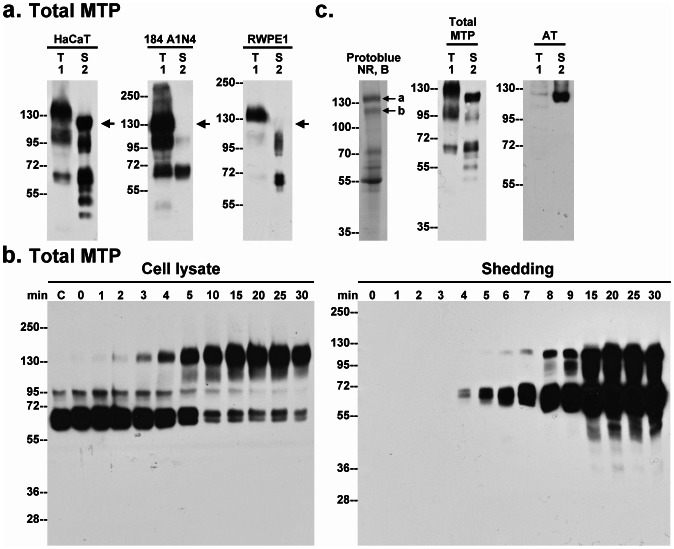
Figure 3. Identification of AT as a component of the novel 110-kDa matriptase complex.
a. Human keratinocytes (HaCaT), mammary (184 A1N4) and prostate (RWPE1) epithelial cells were incubated with a pH 6.0 buffer to induce matriptase activation. The conditioned buffers (S) and the total cell lysates (T) were analyzed by immunoblot for total matriptase. Arrow indicates the 110-kDa matriptase complex. b. HaCaT cells were incubated with a pH 6.0 buffer for the indicated times (minutes). The cell lysates and the conditioned buffers were subjected to immunoblot analyses for total matriptase. c. Left panel: Purified 110-kDa matriptase complex was analyzed by SDS-PAGE under non-reducing and boiled conditions (NR, B). The protein bands were visualized by staining with ProtoBlue Safe. The protein bands a and b, as indicated, were subjected to protein identification by MS/MS (see Fig. S2 for sequence data). Middle and right panels: HaCaT cells, pretreated with human serum, were induced to activate matriptase by pH 6.0 treatment. Cell lysates and the concentrated conditioned buffers were subjected to immunoblot analyses by immunoblot for total matriptase (Total MTP) and AT (AT).

