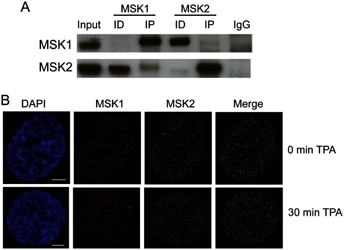
Figure 1. MSK1 and MSK2 are in different complexes.
A. MCF-7 cell lysate (500 µg) was incubated with anti-MSK1 (rabbit, Sigma; 4 µg) or anti-MSK2 (rabbit, Invitrogen; 2 µg) antibodies. The immunoprecipitates (IP) and equivalent volumes of lysate (Input), immunodepleted (ID) fractions, and IgG control (IgG), corresponding to 50 µg of lysate, were loaded onto SDS-8% polyacrylamide gels, transferred to nitrocellulose membranes, and immunochemically stained with anti-MSK1 or anti-MSK2 antibodies. B. MCF-7 cells grown on coverslips were serum starved and then treated with or without TPA as described in Materials and Methods . The cells were fixed and immunostained with antibodies against MSK1 and MSK2, and co-stained with DAPI. Spatial distribution was visualized by fluorescence microscopy and image deconvolution was done by AxioVision software. Yellow signal in the merged images indicates colocalization. Bar, 5 µm.

