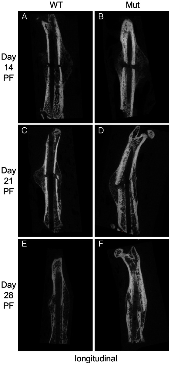
Figure 2. Representative longitudinal µCT sections of the fracture.

The Pten mutants had more bone formation at the proximal and distal ends of the fracture callus at each time point.

The Pten mutants had more bone formation at the proximal and distal ends of the fracture callus at each time point.