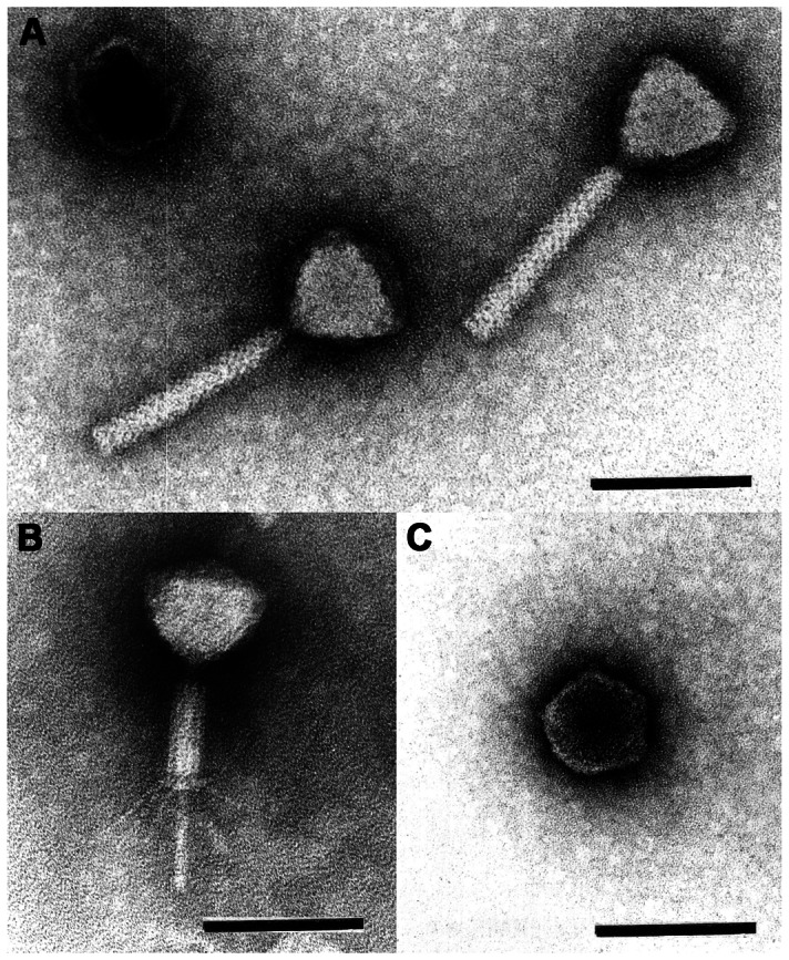
Figure 1. Electron micrographs of purified PaP1 phage particles.

A shows two PaP1 particles with uncontracted tails and an empty head (uranyl acetate). B shows the contracted tail with straight tail fibers (phosphotungstate). C shows a pentagonal head (uranyl acetate). The scale bar represents 100 nm. These micrographs were taken by Hans-Wolfgang Ackermann, School of Medicine, Laval University, Quebec, Canada.
