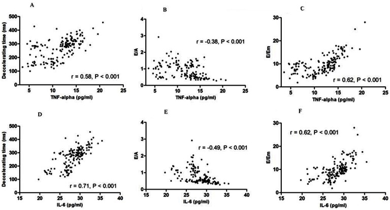
Figure 3. Correlation between interleukin-6 (IL-6), tissue necrosis factor-alpha (TNF-α), and echocardiographic diastolic function parameters in PD patients.
(A) TNF-α and mitral valve ejection flow deceleration time (DT, r = 0.58, P<0.001); (B) TNF-α and the ratio of mitral valve ejection flow (E) divided by mitral valve atrium flow (A, r = −0.38, P<0.001); (C) TNF-α and the ratio of E divided by early diastolic lengthening velocities in tissue Doppler imaging (Em, r = 0.62, P<0.001); (D) IL-6 and DT (r = 0.71, P<0.001); (E) IL-6 and E/A (r = −0.49, P<0.001); and (F) IL-6 and E/Em (r = 0.62, P<0.001).

